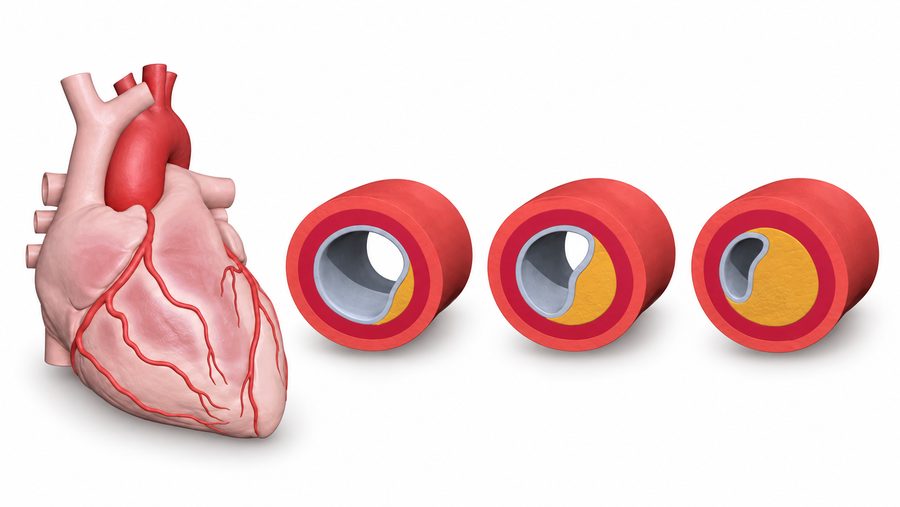
Why plaque and narrowing are different questions
Atherosclerotic plaque builds up within the artery wall. The amount and composition of plaque and the degree of narrowing describe different aspects of the finding. A vessel can contain plaque without a severe narrowing. In HLC care, this information is considered with cholesterol, blood pressure, smoking, diabetes, symptoms and family history.
What CT and additional analysis can show
CT angiography uses X-rays and an intravenous iodine-containing contrast agent to image the coronary arteries. An additional quantitative analysis, when ordered, can organize the findings by artery and describe plaque volume, calcified and non-calcified components, and narrowing. The radiological interpretation remains essential.
This is different from a coronary calcium score, which is a non-contrast CT measure of calcified plaque. Neither test predicts with certainty whether or when an individual will have a heart attack. The clinician chooses the examination that best answers the clinical question.
Before and during the examination
The team reviews kidney function, contrast reactions, medicines, possible pregnancy and heart rhythm. Heart-rate control or other preparation may be needed. During scanning you lie on the CT table, receive contrast through a vein and follow short breath-hold instructions. Follow the imaging centre’s preparation rather than changing medicines yourself.
How the findings guide prevention
The useful output is a clinical decision: whether prevention needs to be strengthened, a symptom requires further investigation or an existing plan should continue. The care team explains which findings are actionable, who will follow them and whether another test is needed. A routine scan is not the right route for sudden chest pain or other urgent symptoms.