Why images and measurements belong together
Brain health is broader than any scan or score. In HLC care, imaging is considered alongside symptoms, medical and family history, vascular risk, sleep and cognitive concerns. Its value is in answering a defined question and identifying whether a finding needs further assessment.
What the two reports describe
The radiologist examines the image sequences and describes visible anatomy, findings and limitations. Quantitative software can then estimate the volume or thickness of selected structures and compare them with a reference group. These calculated comparisons complement the image review; they do not replace it.
An extended analysis may include the hippocampus, cortical regions, white matter and other structures. The exact regions and analysis version vary. A percentile tells you where a measurement falls in that reference group; it is not a percentage of brain function or a measure of how much brain is “left”.
The scan and preparation
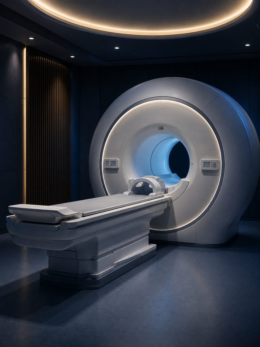
You lie still on a table that moves into the MRI scanner. The equipment makes loud sounds, so hearing protection is provided. Before booking, disclose implants, electronic devices, possible metal fragments, pregnancy, kidney problems and claustrophobia. The imaging team checks safety and explains any preparation for the specific protocol.
What an age estimate does not tell us
Some analyses produce a model-based brain-age estimate. It is not a diagnosis of dementia, a prediction of an individual’s future memory, or proof of rejuvenation after an intervention. Image quality, scanner settings, reference data and software version can affect the output.
If follow-up imaging is useful, the team should make the acquisition and analysis comparable. The practical discussion focuses on clinically relevant findings and modifiable health priorities, including vascular risk, exercise and sleep, rather than trying to optimize one software score.